
Dendritic Cells for Cancer
Dendritic Cells for Cancer:
Dendritic cells are antigen-presenting cells (APCs) which play a critical role in the regulation of the adaptive immune response. Dendritic cells (DCs) are unique APCs and have been referred to as “professional” APCs, since the principal function of DCs is to present antigens, and because only DCs have the ability to induce a primary immune response in resting naive T lymphocytes. DCs were first described by Ralph Steinman nearly thirty years ago. He found a population of striking dendritic-shaped cells in the spleen. Shortly thereafter it became clear that DCs existed in all lymphoid and most non-lymphoid tissues.
Function of Dendritic Cells:
The function of DCs falls broadly into three categories, each of which involve antigen presentation. The first category of DCs function is antigen presentation and activation of T cells. The second category of DC function is not as well established, but it has been suggested that a different class of DCs exist with the function of inducing and maintaining immune tolerance. The third category of DCs, known as follicular DCs, appear to work to maintain immune memory in tandem with B cells.
Development of Dendritic Cells:
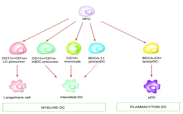
Dendritic Cell Generation:
DCs can be generated by culturing CD34+ cells in the presence of various cytokines. One approach which has been taken involves depleting the CD34+ cells of differentiated precursors and then culturing the cells in the presence of GM-CSF and IL-4 ± TNF-α. CD34+ cells can be obtained from bone marrow, cord blood or G-CSF mobilized peripheral blood. Another approach is to generate DC-like cells by culturing CD14+ monocyte-enriched PBMC. In the presence of GM-CSF and IL-4, these cultures give rise to large numbers of DC like cells.
Working of Dendritic Cells:
Research demonstrate that DCs can capture tumour antigens that are released from tumour cells, either alive or dying, and cross-present these antigens to T cells in tumour-draining lymph nodes. This results in the generation of tumour-specific CTLs that contribute to tumour rejection Thus, DCs represent important targets for therapeutic interventions in cancer.
DCs are found in most tumours in humans and DCs can sample tumour antigens through the capture of dying tumour cells and through the ‘nibbling’ of live tumour cells.
Dying tumour cells mobilize at least three types of signal when interacting with DCs and other phagocytes, including ‘find me’, ‘eat me’ and ‘do not eat me’ signals .There are currently four sets of molecules that are known to be released by apoptotic cells that function as ‘find me’ signals: lipid lysophosphatidylcholine (LPC), sphingosine 1-phosphate (S1P), CX3CL1 (also known as fractalkine), and the nucleotides ATP and UTP. The eat me signals are membrane-bound and serve as markers for phagocytes for recognizing and internalizing dying cells. These signals include phosphatidylserine, alterations in cell-surface charge, αvβ5 integrin and CD36. The eat me signals also include molecules such as milk fat globule-EGF factor 8 (MFG-E8; also known as lactadherin), which bridge the phosphatidylserine of apoptotic cells with the integrin αvβ3 of DCs. Opsonizing antibodies can promote the capture of dying tumour cells through Fc receptors and complement component receptors that are expressed on DCs. The do not eat me signals serve as negative regulators for the capture of cancer cells by DCs and other phagocytes. These signals include lactoferrin and CD47, the interaction of which with signal-regulatory protein-α (SIRPα; also known as SHPS1) on phagocytes provides inhibitory signals that prevent phagocytosis. Accordingly, combining a CD47-blocking antibody with rituximab (a CD20 antibody that depletes B cells) results in enhanced phagocytosis of dead lymphoma cells and improved tumour eradication.
How to Find Us
Address
CTS-1420 Near Shaniwarwada, Kasba peth, Pune -411011
Phone
+91-9511111222
+91-9011111222
admin@universal-hospital.com
anantbagul@yahoo.com